Sight is a remarkable gift. It allows to experience the world in a totally immersive way. But even more remarkable is the complexity behind how the eye works, how it sends information to the brain, and how the brain processes that information.
Step 1: Reception of Photons
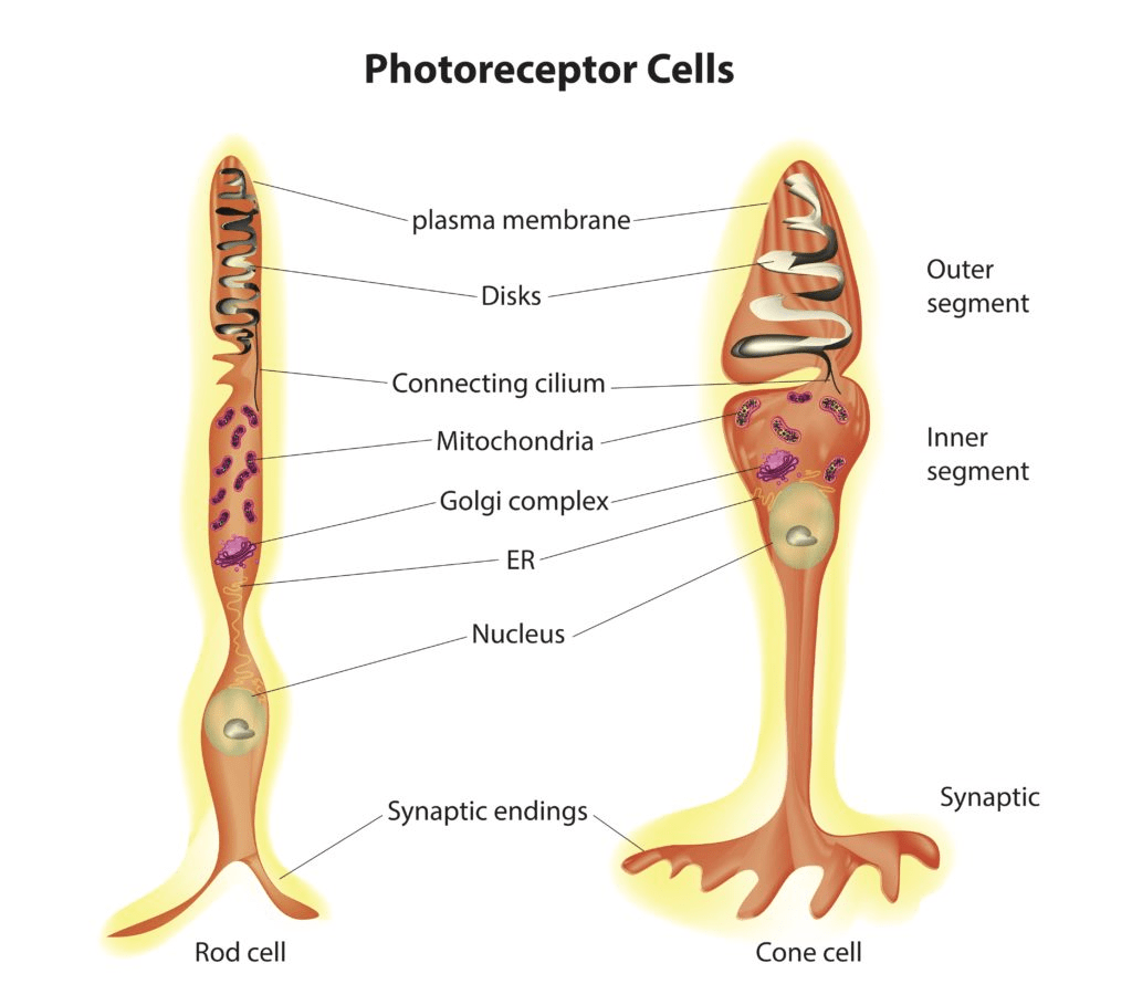
The eye is made of up 6 different types of cells. Two types in particular specialize in taking in photons, and their respective colors. Rod cells (shown below) make up 95% of the photoreceptors in our eyes. These cells are very good at working under low light conditions, as they take in light in shades of grey. However, rod cells have a slow response speed, and cannot contrast between two shades of colors very well, since they only see in black and white. Furthermore, rods cells have a low spacial acuity, or in other terms, cannot see in very fine detail.
Cone cells on the other hand focus more on absorbing color, and picking up all the details that rod cells missed. There are three types of cone cells; Red, Green, and Blue. Red cone cells account for 64% of all cone cells, Green account for 32% and Blue account for 2%. And though cone cells only make up 5% of the total number of cells in the eye, they are vital to our ability to see. Cone cells increase our spacial acuity, and have high reactivity. Due to this ability, many cone cells are concentrated in an area of the retina, known as the fovea. The fovea is the area with the most visual acuity. Cone cells also allow us to see in color!
But how do these cells “see”? Well this process, known as photoreception, starts with the cellular organelle, Disks. The disks are the ones who absorb the photons, and convert them into specific electrical impulses. These electrical signals are then transferred throughout the cell to the synaptic endings. An important detail to notice is the number of mitochondria in each cell. Since rod and cone cells require much energy, they contain many mitochondria in comparison with other cells.
Step 2: Tranmission of Electrical Signals to the Brain
Electrical signals are transferred through the cone/rod cells to the synaptic endings. From here what happens? These synaptic endings connect to another type of cell, known as bipolar cells. Rod cells connect to rod bipolar cells while cone cells connect to cone bipolar cells. This transfer of electrical signals is regulated by Horizontal Cells.
After receiving signals from the photoreceptors, these bipolar cells transmit these signals to Amacrine Cells. These Amacrine cells transmit the electrical signal to the Retinal Ganglion Cells. This whole process is shown below.

Step 3: Understanding and Decoding the Electrical signals
Electrical signals travel along the retinal ganglion cells through a optic nerve. Imagine an optic nerve as a pathway, or an electrical cord for electrical signals. These optic nerves all converge at the Lateral Geniculate Nucleus (LGN). This LGN, located in the thalamus, filters out unnecessary visual information. From here, the visual information (electrical signals) are taken to the visual cortex. The Visual Cortex analyzes the information in layers.
In the primary visual cortex (V1), neurons are specialized for detecting various features such as edges, orientation, color, and motion. In further levels such as V2, V3, and V4, neurons analyze more complex features, such as object recognition, faces, and spatial relationships.
Overall, in the visual cortex, neurons are organized into clusters known as orientation columns. These allow us to visual objects in different orientations and realize they are the same object.
Step 4: Processing the Visual Cortex’s Analysis
Information from the visual cortex is processed in two main ways, the dorsal stream and the ventral stream.
- The dorsal stream focuses on where an object is located in 3d space, allowing us to catch tennis balls, avoid running into walls, and understand the inverse relation between “how far away is something” and its apparent size.
- The ventral stream allows us to identify what an object is. This helps us with face recognition, and day-to-day differenciation between two distinct objects.
A nice way to remember the difference between the dorsal stream and the ventral stream is that the dorsal stream processes visual information for the purpose of “action”, while the ventral stream processes information for the purpose of “perception”.

Leave a comment