Nanotech. I know it best as the material Tony Stark used to engineer his final Iron Man suit (the one that he died in). However, recently it has slowly emerged into the field of neuroscience. Let’s explore how nanotech works in neuroscience!
The Basics
Firstly, let’s talk about what nanotech is. Nanotechnology, short form: nanotech, is the engineering and manipulation of small materials (1-100 nanometers in size). For reference, an human hair is 100,000 nanometers wide. So why are these small particles so innovative in neuroscience?
Firstly, they can cross the blood-brain barrier (BBB). Since, these particles are so small, they can passively cross the blood-brain barrier. They can also target receptors on BBB cells, thus enabling them to cross the blood-brain barrier. Since, about 5% of a dose of a drug only crosses the blood-brain barrier, using nanotech to carry the drug directly into the brain through the BBB could help increase fight diseases such as Alzhiemer’s and epilepsy.
Secondly, nanotech is helping with imaging what happens inside the brain. Scientists are working on quantum dots (semiconductor particles a few nanometers in size) to track individual neurons. Furthermore, they are using nanoparticle based contrast agents, which are made of magnetic nanoparticles, to enhance contrast in MRI imaging.
Lastly, nanotech is being used for neural tissue engineering. Basically, nanoparticles are arranged into nanofibers that shape scaffolds. These scaffolds are able guide axon growth, kind of similar to using a stick to make a tree grow straight. Additionally, carbon nanotubes can be used to increase cell adhesion (process in which cells interact and/or attach to neighboring cells).
Crossing the Blood-Brain Barrier
Nanoparticles can potentially cross the blood-brain barrier passively if they are small enough. However there are other methods as well. For example, nanoparticles can be functionalized (having organic or inorganic molecules attached) with ligands. These ligands would bind to certain receptors on the blood-brain barrier endothelial cells, thus causing transcytosis (a process of transporting materials through a cell by putting them in a membrane-coated small sac or vesicle). Another way that nanoparticles can be used to cross the BBB is via cell mediated transcytosis. The nanoparticle acts as part of a immune cell, thus naturally crossing the BBB. This sometimes is casually known as the “Trojan Horse” method.
Now after crossing the blood-brain barrier, nanoparticles typically do one of two functions. Firstly they deliver drugs, being able to target certain areas of the brain. Secondly they deliver DNA or RNA for gene therapy. A really cool application of nanotech gene therapy is the use of PEGylated liposomes (liposomes coated with polyethylene glycol polymers) to deliver siRNA for Huntington gene silencing, thus helping with Huntington’s disease.
Imaging Using Nanotech
Before understanding how nanotech has enhanced MRI imaging, let’s briefly go over how an MRI works.
- A large magnet in the MRI machine creates a strong magnetic field. This magnet aligns all the protons in every water molecule in the area the machine is scanning.
- A radio wave is sent through the patient which causes all the protons to spin out of alignment.
- When the radio wave is turn on, the protons align back with the magnetic field, thus releasing energy.
- The MRI machine detects this energy and turns it into signals
- A computer turns the signals into digital images.
- Different strength of signals can translate to different shades of grey, indicating different types of tissue.
Now what nanotech does is amplify contrast. The nanoparticles, commonly super paramagnetic iron oxide nanoparticles (SPIONS), interact with the radio waves to produce stronger disruptions. Thus, when the radio waves are turned off, the different amounts of energy each proton sends back varies much more, allowing researching to identify smaller more local structures they couldn’t see with normal MRI.
Another extremely common imaging technique relying on nanotechnology is Atomic Force Microscopy (AFM). AFM relies on the use of a probe tip, typically less than 10 nanometers in radius. This probe tip measure atomic forces, thus creating atomic-scale models of whatever sample the probe tip is measuring. Functionalized probe tips also can provide chemical information about the sample.
Neural Tissue Engineering
Nanoparticles are used to create scaffolds that act as the extracellular matrix (ECM) of neural tissue. The ECM is a 3D network of proteins and carbohydrates that surrounds brain cells that provides structural support for neurons. By mimicking the ECM, nanoparticle scaffolds are able to guide axons regeneration. Typically these scaffolds also include carbon nanotubes as well as graphene in order to improve electrical conductivity, so that the axon can send out messages. A welcome side-effect of using these nano-materials is that they form tight bonds with neuronal membranes, thus creating message shortcuts between different parts to the neuron, helping the neuron be more efficient.
Nanoparticles also are affecting neuron behavior. In fact neurons grown on nanostructure silicon substrates are 3 times more efficient at conducting information compared to neurons grown on flat surfaces. Furthermore, the topography and chemical properties of the nanostructure can affect whether the cell becomes a glia or a neuron.
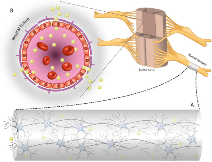
Lastly, nano-engineered conduits are being made to connect large bridge gaps. Similar to scaffolds, these provide support to regrow nerves that were lost or damaged in peripheral nerve injuries. However, these conduits are typically a combination of macro and nano technologies as they support much more mass. Note: A picture of a neural conduit is shown below supporting a nerve located in the spinal cord


Leave a comment