Glial Cells. You’ve probably never heard of them. I know that until writing this article, I didn’t either. A quick google search shows that they work in correlation to neurons. But what exactly are glial cells? How do they work? And what is their purpose?
Types of Glial Cells
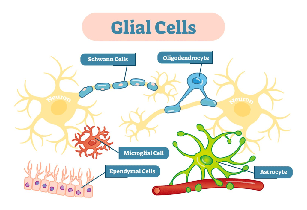
There are five types of glial cells; Astrocytes, Oligodendrocyte, Microglial Cells, Schwann Cells, and Ependymal Cells.
Astrocytes: Astrocytes are in the shape of a star. Well not really, but the do have a central cell body and several protrusions branching off of that central cell body. These cells are located in the CNS (Central Nervous System). Astrocytes have several important functions. First of all, they maintain the nervous tissue structure. They also control ion and nutrient concentrations outside of the cell, and block certain substances found in the bloodstream from entering the brain (blood-brain barrier). Lastly, astrocytes aid in the repair process after brain injury/damage.
Oligodendrocyte: Oligodendrocytes are designed for one purpose only: providing myelin to axons. Thus, oligodendrocytes have these protrusions that then wrap around axons and provide axons with their needed myelin. Since oligodendrocytes have multiple of these protrusions, they can provide myelin to several neurons’ axons at the same time. This way of connecting neurons (just mechanically) helps provide structural integrity to the brain.
Microglial Cells: Microglia are small cells that account for 10% of all the cells in the CNS. They do regulate synaptic transmissions and sculpt neuronal synapses but their primary function occurs after brain damage, or after a concussion. Microglia are crucial for CNS tissue repair. They remove microbes, dead cells and protein aggregates (misfolded proteins). Microglia are also the primary producer of soluble factors such as chemoattractants, cytokines, and neurotropic factors that help with immune response and tissue repair in the CNS.
Schwann Cells: Schwann cells are almost exactly like oligodendrocytes. They also provide myelin to the axons of neurons. The only difference is that Schwann cells are located in the PNS (Periphery Nervous System).
Ependymal Cells: These column-like cells are found in ventricles (fluid filled cavities of the brain), and in the spine. Ependymal cells produce CSF (cerebrospinal fluid). They regulate movement of nutrients in CSF, monitor the blood-CSF barrier, and circulate CSF.
Structure of Glial Cells:
Important Points to Notice:
- Every cell has a small circle; That circle refers to the nucleus of the cell where the DNA is kept.
- All types of glial cells include the protrusions that help them interact with neurons and/or CSF.
- Both schwann cells and oligodendrocytes have protrusions that wrap around axons.
- Ependymal cells, being the only glia that don’t interact much with neurons have much fewer protrusions than all other types of glial cells.
- All glial cells are smaller than neurons, yet astrocytes are close to the size of neurons.
Conclusions:
- In regards to the CNS, the astrocyte and oligodendrocyte work 24/7 and therefore, are much bigger than all the other cells. The microglial cells, who only really have to work in case of brain injury/damage are the smallest glial cell in the CNS.
- In regards to the PNS, the schwann cells also work 24/7 yet aren’t as large as the oligodendrocyte.
- Regarding the spine, Ependymal cells kind of do it all, but due to the structure of the spine, they have evolved in a different way than all other glial cells.

Leave a reply to Multiple Sclerosis: How the Brain Fights Itself – Behind the Brain Cancel reply